

Mark Nicolas · September 16, 2026
The Cerebellum and the Timing of Reward
A new Nature Neuroscience study shows cerebellar circuits tracking when dopamine reward is expected, registering when it arrives, and helping the brain connect behavior with what happens next.
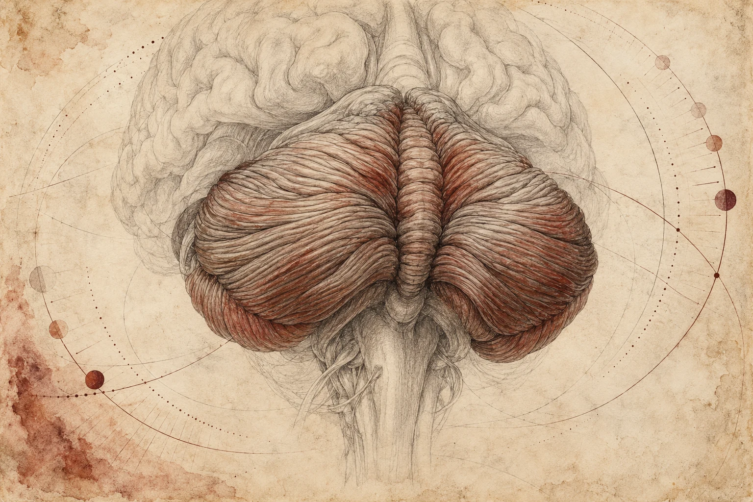
Most people who learned the reward system in a psychology or neuroscience course probably did not spend much time talking about the cerebellum. The usual reward system map runs through the ventral tegmental area, nucleus accumbens, striatum, prefrontal cortex, amygdala, and hippocampus. The cerebellum generally enters the conversation somewhere else, usually through movement, coordination, prediction, and timing. A growing body of research is making that separation harder to maintain.
Benjamin Filio and colleagues (2026) recently published an experiment in Nature Neuroscience examining what the cerebellum is doing while an animal expects a dopamine related reward. The question sounds fairly specific. The way they approached it opens a much larger conversation about how the brain connects an action with an outcome that has not happened yet.
What they were trying to separate
Researchers have known for years that cerebellar neurons respond during reward related behavior (Wagner et al., 2017). Interpreting those signals has always been difficult because natural rewards usually involve movement. Give a thirsty mouse water and it has to lick. Give an animal food and it has to approach it, manipulate it, chew, and swallow. Social rewards come with their own movements and sensory activity. The cerebellum is heavily involved in coordinating movement and predicting its consequences. When cerebellar activity appears around food or water, part of the signal could be related to reward and part could reflect all of the behavior required to obtain and consume it.
Filio and colleagues built an experiment that gave them a cleaner look at the reward side of the problem. Mice learned to push a robotic handle. After completing the movement, they waited before receiving stimulation of reward circuitry inside the brain. One version of the experiment used optogenetic stimulation of dopamine neurons in the ventral tegmental area. Optogenetics uses light to activate neurons that have been genetically made sensitive to it. Another electrically stimulated the medial forebrain bundle, a collection of nerve fibers connecting several forebrain and midbrain regions. Those manipulations are not biologically identical. Ventral tegmental area stimulation allowed more selective engagement of dopamine neurons. Medial forebrain bundle stimulation produces a powerful rewarding effect while recruiting dopamine related and non dopamine fibers.
The important feature of the experiment was the delay. Once the animal finished pushing the handle, there was nothing else it had to do to consume the reward. The mouse pushed. Then it waited. That waiting period gave the researchers a window into what the cerebellum was doing between an action and the consequence that the animal had learned would follow it.
What the cerebellum did while the mouse waited
Many cerebellar granule cells, small neurons that relay incoming information within the cerebellum, began changing their activity during that delay (Filio et al., 2026). Some gradually became more active. Others progressively became less active.
Then the researchers changed the amount of time before the reward arrived. When the animals expected the reward after one second, the neural activity unfolded across roughly that interval. After the animals were retrained with a two second delay, a subset of the same activity patterns stretched across the longer period. The neural dynamics adjusted to the expected timing of the outcome. That gets us beyond describing these neurons as generally “reward responsive.” They were carrying information through time.
The reward omission trials make the pattern even more interesting. On some trials, the expected reward never arrived. The granule cell activity continued beyond the point where the animal had learned the reward should appear. When the reward arrived normally, those anticipatory patterns terminated much more quickly. The simplest way to explain what the animal was doing during those omission trials is that it was still waiting. Apparently, part of the cerebellum was too.
The researchers also tested whether the signal depended on the animal making the original movement. They trained another group of mice in a passive anticipation task. An auditory cue predicted stimulation one second later. The mouse did not have to push the handle. Granule cell activity still ramped during the delay. That makes movement an increasingly poor explanation for the signal. These cells could carry the temporal expectation of an upcoming reward even when there was no instrumental action to bridge to it.
The signal that arrived with the reward
The climbing fibers carried a different piece of the process. These inputs reach the cerebellum from the inferior olive, a structure in the brainstem. Granule cells showed activity while the animal was waiting. Many climbing fibers responded immediately after the dopamine related reward arrived. Those climbing fiber responses were already widespread on the first day of training (Filio et al., 2026). That timing gives them a plausible role as an instructive signal while the animal learns the relationship between what it did and what happened afterward.
Put the two signals together and a useful computational picture starts to form. One cerebellar input system can preserve information through the waiting period. Another provides a strong signal when the expected outcome arrives. The cerebellum now has information about the behavior, the expected timing of its consequence, and the arrival of that consequence within the same learning architecture.
The authors describe the granule cell patterns as a temporal basis set. The terminology sounds more complicated than the underlying problem. Imagine pressing a button and receiving a reward two seconds later. Some part of the nervous system has to maintain enough information about the button press for the reward two seconds later to be associated with it. Otherwise the brain faces what computational neuroscience calls a credit assignment problem. The outcome occurred. What should receive credit for causing it? The temporal pattern distributed across these granule cells may help bridge that interval until the outcome signal arrives.
The causal experiments
Filio and colleagues (2026) also manipulated the circuitry directly. They activated climbing fibers and asked whether animals could learn from that signal itself. Naive mice learned to perform the action for delayed climbing fiber stimulation. The resulting behavior was more moderate than the behavior produced by the main dopamine related reward conditions, but the pathway was capable of reinforcing a new operant behavior.
The investigators then approached the system from the other direction. They disrupted granule cell activity during the delay between the action and the expected reward. Learning deteriorated. Some of that deficit remained during the first washout session after the inhibition was removed, providing evidence that disrupting the delay period had interfered with acquisition of the learned behavior and had effects extending beyond the immediate manipulation.
That moves the paper beyond observing cerebellar activity that happens to occur around reward. These signals are participating in the learning process.
Adding time to the way we think about reward
I keep coming back to the temporal part of this paper. Reward is often discussed in terms of levels and magnitudes. How much dopamine was released? How strongly did a cue activate a region? How large was the BOLD response, the blood oxygen level dependent signal used in functional MRI? How much did connectivity change?
Those measurements give us useful information. A reward process also has a trajectory. A cue is encountered. An outcome becomes expected. Motivation develops around that expectation. Behavior is organized. Time passes. The expected outcome either arrives, arrives late, or never appears. The nervous system then has to update whatever prediction produced the behavior in the first place.
Berridge, Robinson, and Aldridge (2009) separated reward into related processes involving wanting, liking, and learning years ago. That framework helped move reward science away from the idea that dopamine could be treated as a straightforward measure of pleasure.
The Filio paper puts time directly into the architecture. Expectation begins somewhere. It develops at some rate. It persists for some period. It changes when the expected timing changes. It continues when an expected outcome fails to arrive. Eventually it resolves. Those dynamics may contain information that disappears when the entire response gets reduced to one number.
Why the cerebellum keeps showing up
There is another reason this paper caught my attention. The cerebellum has been appearing with surprising regularity in psychedelic neuroscience. We do not yet have a settled explanation for its role in the psychedelic state. The pattern itself is becoming difficult to dismiss as an incidental finding.
In 2024, Joshua Siegel and colleagues used precision functional mapping to examine psilocybin induced changes in the human brain. Some of the largest subcortical functional connectivity changes appeared in the thalamus, basal ganglia, hippocampus, and cerebellum. Within the cerebellum, the largest effects appeared in regions functionally connected with the default mode network, a collection of regions associated with internally directed thought (Siegel et al., 2024). Functional connectivity describes how signals from different regions fluctuate together; it does not by itself establish a direct anatomical connection.
A broader meta analysis published the same year by Kenneth Shinozuka and colleagues looked across the psychedelic neuroimaging literature. Their analysis found increased connectivity from the cerebellum to the cortex among the significant subcortical findings. That paper also deserves an important qualification. The authors rated confidence in the functional connectivity literature as low because of methodological limitations and risk of bias across the available studies (Shinozuka et al., 2024).
The signal kept appearing as larger datasets became available. In 2026, Manesh Girn and a large international group combined 11 independent resting state fMRI datasets covering psilocybin, LSD, mescaline, DMT, and ayahuasca. The study included 267 unique participants and more than 500 scanning sessions analyzed through a common pipeline. The cerebellum again appeared in the architecture of psychedelic induced network change. The authors found altered cerebellar coupling with sensorimotor networks alongside changes involving structures such as the thalamus, caudate, and putamen (Girn et al., 2026). These networks help organize sensation and movement. The strength and certainty of the connectivity effects varied across drugs, network pairs, and preprocessing choices.
Then mescaline produced another cerebellar finding from a very different experimental direction. Noah Cavallaro and colleagues used awake rat fMRI and reported pronounced cerebellar effects after mescaline. They observed cerebellar BOLD suppression together with increased functional connectivity involving the cerebellum, hippocampus, thalamus, somatosensory cortex, and midbrain.
They proposed that altered cerebellar processing could disrupt sensory filtering during the psychedelic state (Cavallaro et al., 2026). That last part is a proposed mechanism from an animal study. It gives us a question to test, not a conclusion about what is happening in humans.
Across these studies, though, the cerebellum keeps reappearing. The Filio paper gives that recurring neuroimaging observation more context. Granule cells can carry the expected timing of reward. Climbing fibers can signal the arrival of an outcome. Changing temporal expectations changes the neural dynamics. Disrupting those dynamics changes learning.
The psychedelic imaging literature usually cannot tell us what a cerebellar circuit is computing. It can tell us that relationships between regions have changed. Once we know that the cerebellum is capable of computations involving timing, prediction, sensory processing, expected outcomes, and learning, altered cerebellar connectivity during a psychedelic state becomes a more interesting observation.
I want to know which of those computations are changing. Does a psychedelic alter the temporal precision of prediction? Does it change how expected and unexpected sensory information is weighted? Does it change the gain on prediction error signals? Does it affect how long an expectation remains active after an expected outcome fails to appear? Does altered cerebellar communication contribute to the unusual relationship between sensation, expectation, salience, and meaning that occurs during a psychedelic experience? We do not have answers to those questions yet. They are worth asking.
How this connects with reward fidelity
The same temporal logic has implications for the way I have been thinking about reward system fidelity. My interest has been in whether a dysfunctional reward system assigns motivational value appropriately and whether that function can recover, as I proposed in my published reward system paper (Nicolas, 2025). A drug cue, traumatic cue, food reward, social reward, compulsive behavior, and naturally rewarding experience can each recruit motivational learning differently. Measuring how strongly the nervous system responds gives us part of that picture. The Filio study gives us a good experimental reason to also examine the shape of the response across time.
How quickly does expectation develop after a cue? Does the duration of the response match the expected timing of the outcome? What happens when the outcome is delayed? How long does expectation persist when the expected reward never appears? How quickly does the system disengage after the reward arrives? Does the temporal organization generalize appropriately between drug related rewards and natural rewards? What happens to those properties during recovery? Those are measurable questions.
I am not treating this mouse study as validation of Reward Fidelity. What it gives me is an experimental example of why temporal organization may contain information about reward function that amplitude alone cannot capture. A system can generate a large response and still organize that response poorly. Timing, persistence, flexibility, and recovery may tell us something different.
The connection to neural attunement
There is a second connection in the experimental design itself. The researchers changed the reward delay. They omitted the expected reward. They changed the reward. They removed the instrumental action. They activated one pathway. They inhibited another. Each manipulation exposed another property of the system.
That experimental logic is very close to the Rest → Perturbation → Response → Recovery framework I have been developing within Neural Attunement. A resting measurement gives us a state. A perturbation allows us to watch the system behave.
For a reward related probe, I would want to know the baseline state, response latency, response amplitude, gain, propagation, duration of expectation, coupling with other systems, response to omission, and the trajectory back toward baseline. I would also want to know whether repeated perturbations produce appropriate adaptation or whether the same network becomes increasingly rigid, unstable, or context insensitive.
The Filio study was designed to investigate cerebellar reward learning. It was not designed to test Neural Attunement. The useful overlap is methodological. Their results show how much information becomes visible when neural function is examined dynamically across perturbations instead of being summarized from a resting state or single peak response. That is the part I find especially useful.
Where the boundaries are
There are limits to how far any of this can be taken. The Filio experiments were performed in mice. The reward conditions involved controlled direct brain stimulation. Electrical medial forebrain bundle stimulation recruits more than dopamine fibers. The work focused heavily on cerebellar lobule VI, one subdivision of the cerebellum (Filio et al., 2026).
The psychedelic imaging studies use different compounds, methods, analysis pipelines, species, and experimental conditions. Functional connectivity tells us that statistical relationships between signals changed. It does not identify the cellular computation producing the change. The mescaline study was performed in awake rats, so its proposed sensory filtering mechanism should remain a hypothesis when discussing human psychedelic states.
None of these studies demonstrate that psychedelic treatment works by changing cerebellar reward timing. They do give us several converging reasons to start taking the cerebellum more seriously in both reward science and psychedelic neuroscience. The brain has to preserve information about what happened, estimate what should happen next, track when it should happen, recognize the outcome when it arrives, and update its behavior when the prediction fails. The cerebellum appears to be involved in considerably more of that process than the simplified reward diagrams many of us learned would suggest.
For me, the next question is no longer whether the cerebellum belongs in the conversation. It is what information the cerebellum is carrying when these systems are perturbed, and whether the organization of that information changes during recovery.
References
- Berridge, K. C., Robinson, T. E., & Aldridge, J. W. (2009). Dissecting components of reward: “Liking,” “wanting,” and learning. Current Opinion in Pharmacology, 9(1), 65–73. https://doi.org/10.1016/j.coph.2008.12.014
- Cavallaro, N., Rai, P., Akins, D., Soltanpour, S., Nasseef, M. T., Ortiz, R. J., Utama, R., Cody, C. R., Mistry, A., Brenhouse, H. C., Kulkarni, P. P., & Ferris, C. F. (2026). Mescaline alters cerebellar function, global connectivity, and frequency-selective acoustic gating: A BOLD fMRI study in awake rats. Neuroscience Bulletin. Advance online publication. https://doi.org/10.1007/s12264-026-01632-3
- Filio, B. A., Otchere, A., Srinivasan, S., Thota, S., Drake, L., Ramos, L., Maurus, P., & Wagner, M. J. (2026). Predictive and instructive cerebellar encoding of dopamine reward drives motivated behavior. Nature Neuroscience. Advance online publication. https://doi.org/10.1038/s41593-026-02449-z
- Girn, M., Doss, M. K., Roseman, L., Preller, K. H., Palhano-Fontes, F., Pasquini, L., Barrett, F. S., Mallaroni, P., Mason, N. L., Timmermann, C., McCulloch, D. E., Fisher, P. M., Winston, B. S., Moujaes, F., Muller, F., Liechti, M. E., Vollenweider, F. X., Ramaekers, J. G., Kuypers, K., … Bzdok, D. (2026). An international mega-analysis of psychedelic drug effects on brain circuit function. Nature Medicine, 32(4), 1543–1554. https://doi.org/10.1038/s41591-026-04287-9
- Nicolas, M. (2025). Ibogaine’s potential role in supporting reward system recovery across diagnostic boundaries. Frontiers in Pharmacology, 16, Article 1744383. https://doi.org/10.3389/fphar.2025.1744383
- Shinozuka, K., Jerotic, K., Mediano, P., Zhao, A. T., Preller, K. H., Carhart-Harris, R., & Kringelbach, M. L. (2024). Synergistic, multi-level understanding of psychedelics: Three systematic reviews and meta-analyses of their pharmacology, neuroimaging and phenomenology. Translational Psychiatry, 14, Article 485. https://doi.org/10.1038/s41398-024-03187-1
- Siegel, J. S., Subramanian, S., Perry, D., Kay, B. P., Gordon, E. M., Laumann, T. O., Reneau, T. R., Metcalf, N. V., Chacko, R. V., Gratton, C., Horan, C., Krimmel, S. R., Shimony, J. S., Schweiger, J. A., Wong, D. F., Bender, D. A., Scheidter, K. M., Whiting, F. I., Padawer-Curry, J. A., … Dosenbach, N. U. F. (2024). Psilocybin desynchronizes the human brain. Nature, 632(8023), 131–138. https://doi.org/10.1038/s41586-024-07624-5
- Wagner, M. J., Kim, T. H., Savall, J., Schnitzer, M. J., & Luo, L. (2017). Cerebellar granule cells encode the expectation of reward. Nature, 544, 96–100. https://doi.org/10.1038/nature21726